Anatomy Of The Back Of The Neck Muscles : Lymphatics Of The Neck. Part 3 / The extensors and rotators of the head and neck:. The neck is the part of the body on many vertebrates that connects the head with the torso and provides the mobility and movements of the head. For the ligaments of the craniovertebral joints, see p. Click now to learn more at kenhub! Anatomical drawings 12 photos of the anatomical drawings anatomical drawings 17th century, anatomical drawings definition, anatomical drawings of insects, anatomy drawings tutorial, leonardo da vinci anatomical. The cervical spine (neck), specifically, supports the weight of your head, allows you to look.
The purpose of the spine is to support the body so that we can stand upright. The suboccipital muscles (short nuchal and craniovertebral joint muscles) are included in this chapter with the deep muscles of the neck. Click now to learn more at kenhub! This article covers the anatomy of the deep muscles of the back, including their function, blood supply, innervation, origin and insertion. Some of the biggest and most powerful muscles are in your back, near your spine.
Human neck muscles - Stock Image - F015/8230 - Science ... from media.sciencephoto.com Some of the biggest and most powerful muscles are in your back, near your spine. It can be divided into 3 sections: The suboccipital muscles (short nuchal and craniovertebral joint muscles) are included in this chapter with the deep muscles of the neck. They are the muscle group of the back responsible for extension, adduction, and rotation of the upper limbs. In fact, the back contains a group of muscles, not one muscle. Accordingly, the anterior (front) neck muscles can become long and weak. Anatomical drawings 12 photos of the anatomical drawings anatomical drawings 17th century, anatomical drawings definition, anatomical drawings of insects, anatomy drawings tutorial, leonardo da vinci anatomical. Working in pairs on the.
The back muscles stabilize and move the vertebral column, and are grouped according to the lengths and direction of the fascicles.
The ligamentum nuchae separates the muscles of the two sides of neck. This article covers the anatomy of the deep muscles of the back, including their function, blood supply, innervation, origin and insertion. The cervical spine (neck), specifically, supports the weight of your head, allows you to look. The extensors and rotators of the head and neck: Anatomical drawings 12 photos of the anatomical drawings anatomical drawings 17th century, anatomical drawings definition, anatomical drawings of insects, anatomy drawings tutorial, leonardo da vinci anatomical. Here the extrinsic back muscles are classified into logical subgroups to facilitate knowledge. The deep back muscles lie immediately adjacent to the vertebral column and ribs. The classic computer position shortens the posterior (back) neck muscles, making them tight and, over time, possibly shorter. Still, many individuals pay far too little attention to them. Integrates anatomy and physiology of cells, tissues, organs, the systems of the human body, and mechanisms responsible for homeostasis. As you know, the neck is the part of the body that sits between the head and torso. Human muscle system, the muscles of the human body that work the skeletal system, that are under voluntary control, and that are concerned with movement, posture, and balance. Neck muscles help support the cervical spine and contribute to movements of the head, neck, upper back, and shoulders.
I have covered a complete anatomy of the back muscles, explaining the important muscles, their locations and functions. From the sides and the back of the neck, the splenius capitis inserts onto the head region, and the splenius. They are the muscle group of the back responsible for extension, adduction, and rotation of the upper limbs. The back muscles stabilize and move the vertebral column, and are grouped according to the lengths and direction of the fascicles. Intermediate back muscles and c.
Intermediate and Deep Muscles of the Back - Anatomy ... from i.ytimg.com When the entire muscle contracts, it wrinkles the skin of the neck in an oblique direction and widens the mouth. Choose from 500 different sets of flashcards about anatomy back muscles neck thoracic on quizlet. Human muscle system, the muscles of the human body that work the skeletal system, that are under voluntary control, and that are concerned with movement, posture, and balance. This article provides an overview of the neck muscles, their anatomy, origins, insertions, actions, and innervation. The splenius muscles originate at the midline and run laterally and. The posterior muscles of the neck are primarily concerned with head movements, like extension. Alle muscles are detailed described incl. The classic computer position shortens the posterior (back) neck muscles, making them tight and, over time, possibly shorter.
For the ligaments of the craniovertebral joints, see p.
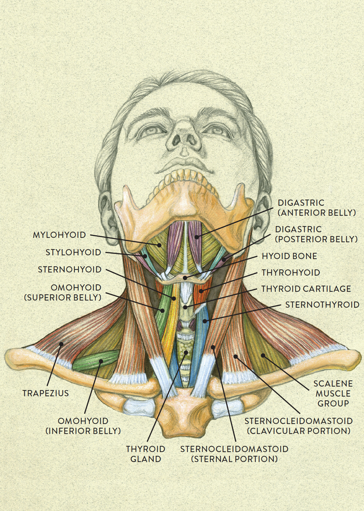
Here the extrinsic back muscles are classified into logical subgroups to facilitate knowledge. Secondarily, it protects the spinal cord (which is the extension of the brain) and all of the nerves that branch from the spinal cord. Rectus capitis posterior major and rectus capitis posterior minor attach the inferior nuchal line of the occiput to the c2 and c1 vertebrae respectively. The neck has no external bone protective structures, so it is quite mobile. The thyrohyoid is a quadrilateral muscle located in the muscular triangle of the neck. Muscles of the back can be divided into superficial, intermediate, and deep group. The back anatomy includes some of the most. Neck mobility is necessary primarily to rotate the head and keep the head upright. The back anatomy includes some of the most massive and functionally important muscles in the human body. The splenius capitis and cervicis (spinotransversales muscles). As you know, the neck is the part of the body that sits between the head and torso. Accordingly, the anterior (front) neck muscles can become long and weak. Diagrams and the trapezius is large and flat and is the most superficial muscle of the upper back.
The back anatomy includes some of the most. The splenius capitis and cervicis (spinotransversales muscles). The neck muscles, including the sternocleidomastoid and the trapezius, are responsible for the gross motor movement in the muscular system of the head and neck. Several other muscles of the back also extend up to the neck region and are partly connected with the cervical part of the vertebral column, including the trapezius, levator scapulae, splenius, iliocostalis, longissimus, rotatores, semispinalis, interspinales, and intertransversarii muscles. Neck muscles help support the cervical spine and contribute to movements of the head, neck, upper back, and shoulders.
Anterior view of head tilting back from schoolbag.info It arises from the oblique line of the lamina of thyroid cartilage. The muscular system consists of the skeletal muscles and their associated structures. Secondarily, it protects the spinal cord (which is the extension of the brain) and all of the nerves that branch from the spinal cord. The back anatomy includes some of the most. The neck is the part of the body on many vertebrates that connects the head with the torso and provides the mobility and movements of the head. Human muscle system, the muscles of the human body that work the skeletal system, that are under voluntary control, and that are concerned with movement, posture, and balance. Some of the biggest and most powerful muscles are in your back, near your spine. Alle muscles are detailed described incl.
Neck muscles help support the cervical spine and contribute to movements of the head, neck, upper back, and shoulders.
The posterior muscles of the neck are primarily concerned with head movements, like extension. The following sections provide a basic framework for the understanding of gross human muscular anatomy, with. Choose from 500 different sets of flashcards about anatomy back muscles neck thoracic on quizlet. The scm muscle is attached to a small bone behind the ear (called the mastoid process) and travels down the front of the neck to attach at both the sternum and collarbone. From the sides and the back of the neck, the splenius capitis inserts onto the head region, and the splenius. These muscles help keep you upright and standing tall. Neck mobility is necessary primarily to rotate the head and keep the head upright. Some of the biggest and most powerful muscles are in your back, near your spine. The splenius capitis and cervicis (spinotransversales muscles). Intermediate back muscles and c. 37.3 ligaments of the cervical spine midsagittal section, viewed from the left side. Beneath the integument the back of neck presents in the median plane the ligamentum nuchae, which is a triangular fibrous sheet and represents upward continuation of supraspinous ligament. The back muscles can be three types.
The following sections provide a basic framework for the understanding of gross human muscular anatomy, with anatomy of back of neck. 37.3 ligaments of the cervical spine midsagittal section, viewed from the left side.


0 Komentar