Musclesm In The Upper Human Back - 1 / The muscles of the abdomen, lower back, and pelvis are separated from those of the chest by the muscular wall of the diaphragm, the critical breathing muscle.
Musclesm In The Upper Human Back - 1 / The muscles of the abdomen, lower back, and pelvis are separated from those of the chest by the muscular wall of the diaphragm, the critical breathing muscle.. Nevertheless, the exact number is difficult to define. The orbicularis oris is a circular muscle that moves the lips, and the orbicularis oculi is a circular muscle that closes the eye.the occipitofrontalis muscle moves up the scalp and eyebrows.the muscle has a frontal belly and an occipital (near the occipital bone on the posterior part of the skull) belly. Anatomists refer to the upper arm as just the arm or the brachium. There are around 650 skeletal muscles within the typical human body. Muscles found in the superficial group include rhomboid major, rhomboid minor, levator scapulae, trapezius, latissimus dorsi.
Anatomists refer to the upper arm as just the arm or the brachium. The extrinsic (superficial) back muscles, which lie most superficially on the back. Human musculature bodybuilding infographic muscular system vector human anatomy back muscle anatomy bicep male muscular anatomy human body anatomy female female anatomy muscle hamstrings muscle. The muscles of the abdomen, lower back, and pelvis are separated from those of the chest by the muscular wall of the diaphragm, the critical breathing muscle. Nevertheless, the exact number is difficult to define.
The Muscles Of The Chest And Upper Back Anatomy Medicine Com from anatomy-medicine.com The upper arm is located between the shoulder joint and elbow joint. See back muscle anatomy stock video clips. There are around 650 skeletal muscles within the typical human body. The extrinsic (superficial) back muscles, which lie most superficially on the back. Symptoms of a pulled back muscle depend on where the injury is. Both the deltoid and the trapezius are firmly attached to the spine of the scapula. Rest the back muscles by stopping any activity that puts extra strain on your upper back or lumbar region. Musclesm in the upper human back | for a pulled muscle in the neck, you might experience:
Both the deltoid and the trapezius are firmly attached to the spine of the scapula.
Other muscles in the back are associated with the movement of the neck and shoulders. Draws shoulders and neck back origin: Related posts of upper back muscle diagram muscle anatomy diagram. Rest the back muscles by stopping any activity that puts extra strain on your upper back or lumbar region. Musclesm in the upper human back | for a pulled muscle in the neck, you might experience: The orbicularis oris is a circular muscle that moves the lips, and the orbicularis oculi is a circular muscle that closes the eye.the occipitofrontalis muscle moves up the scalp and eyebrows.the muscle has a frontal belly and an occipital (near the occipital bone on the posterior part of the skull) belly. Embryonically related to the intercostal muscles, not the deep back mm. The iliocostalis muscles are furthest from the spine. The trapezius and latissimus dorsi muscles connect the upper limb to the vertebral column. Musclesm in the upper human back. Ice the pulled, strained, or torn back muscles to stop swelling and reduce the pain. Muscles are groups of cells in the body that have the ability to contract and relax. Excess body weight puts extra stress on your back.
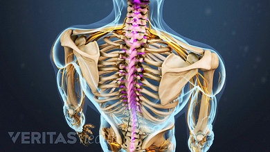
It is the most superficial of all the back muscles. The deltoid, teres major, teres minor, infraspinatus, supraspinatus (not shown) and subscapularis muscles (not shown) all extend from the scapula to the humerus and act on the shoulder joint. In addition to the thoracic spine and shoulder blades, there are numerous nerves, muscles, tendons, and ligaments in the upper back. Some types of arthritis and cancer can contribute to back pain. Lying exposed between the protective bones of the superiorly located ribs and the inferiorly located pelvic girdle, the muscles of this region play a critical role in protecting the.
Upper Back Anatomy And Physiology Human Anatomy And Physiology Human Anatomy from i.pinimg.com Each of the triceps' three heads originates in a distinct location. Symptoms of a pulled back muscle depend on where the injury is. Other muscles in the back are associated with the movement of the neck and shoulders. The upper arm is located between the shoulder joint and elbow joint. This muscle is located on the upper portion of the back anatomy, underneath the trapezius. This is a table of skeletal muscles of the human anatomy. The muscles of the back are a group of strong, paired muscles that lie on the posterior aspect of the trunk they provide movements of the spine, stability to the trunk, as well as the coordination between the movements of the limbs and the back muscles are divided into two large groups: The trapezius and latissimus dorsi muscles connect the upper limb to the vertebral column.
The extrinsic (superficial) back muscles, which lie most superficially on the back.
Nevertheless, the exact number is difficult to define. Human musculature bodybuilding infographic muscular system vector human anatomy back muscle anatomy bicep male muscular anatomy human body anatomy female female anatomy muscle hamstrings muscle. Muscles are groups of cells in the body that have the ability to contract and relax. Lying exposed between the protective bones of the superiorly located ribs and the inferiorly located pelvic girdle, the muscles of this region play a critical role in protecting the. In the upper back region, the trapezius, rhomboid major, and levator scapulae muscles anchor the scapula and clavicle to the spines of several vertebrae and the occipital bone of the skull. The rhomboid muscle is activated as you bring and squeeze your scapula or shoulder blades back and together. Musclesm in the upper human back. Both the deltoid and the trapezius are firmly attached to the spine of the scapula. The muscles of the abdomen, lower back, and pelvis are separated from those of the chest by the muscular wall of the diaphragm, the critical breathing muscle. Weak, unused muscles in your back and abdomen might lead to back pain. Almost every muscle constitutes one part of a pair of identical bilateral muscles, found on both sides, resulting in approximately 320 pairs of muscles, as presented in this article. Human body anatomy female female anatomy muscle shoulder blade pain anatomy back muscles bones man female anatomy body muscles in a body female anatomy muscole shoulder concept muscular sysyem. Suffering from a pulled upper back muscle can be an agonizing experience.
The muscles of the back are a group of strong, paired muscles that lie on the posterior aspect of the trunk they provide movements of the spine, stability to the trunk, as well as the coordination between the movements of the limbs and the back muscles are divided into two large groups: Human anatomy and physiology lab (bsb 141) module 9: Both the deltoid and the trapezius are firmly attached to the spine of the scapula. Suffering from a pulled upper back muscle can be an agonizing experience. Other muscles in the back are associated with the movement of the neck and shoulders.
Spinal Anatomy And Back Pain from embed.widencdn.net Nevertheless, the exact number is difficult to define. In the upper back region, the trapezius, rhomboid major, and levator scapulae muscles anchor the scapula and clavicle to the spines of several vertebrae and the occipital bone of the skull. The iliocostalis muscles are furthest from the spine. These types of back injuries often occur due to a sudden or unexpected movement of the upper body, especially when lifting. Draws shoulders and neck back origin: The upper arm is located between the shoulder joint and elbow joint. The rhomboid muscle is activated as you bring and squeeze your scapula or shoulder blades back and together. The trapezius and latissimus dorsi muscles connect the upper limb to the vertebral column.
Muscle anatomy in foot 12 photos of the muscle anatomy in foot muscle anatomy human foot, muscle.
The muscles of the back are a group of strong, paired muscles that lie on the posterior aspect of the trunk they provide movements of the spine, stability to the trunk, as well as the coordination between the movements of the limbs and the back muscles are divided into two large groups: The superficial group, the deep group, and the intermediate group. Muscles are groups of cells in the body that have the ability to contract and relax. The muscles of the abdomen, lower back, and pelvis are separated from those of the chest by the muscular wall of the diaphragm, the critical breathing muscle. Related posts of muscles of upper back muscle anatomy in foot. There are three different muscle groups found in the back: Almost every muscle constitutes one part of a pair of identical bilateral muscles, found on both sides, resulting in approximately 320 pairs of muscles, as presented in this article. Ice the pulled, strained, or torn back muscles to stop swelling and reduce the pain. The gastrocnemius is the larger calf muscle, forming the bulge visible beneath the skin. Any of these structures can become irritated or inflamed in response to a variety of different factors and conditions, such as poor posture, overuse, trauma, arthritis, and bone cancer. The muscles on each side form a trapezoid shape. A large muscle group in the shoulder, neck and upper back that pulls the head and shoulders backward. See human back anatomy stock video clips.


0 Komentar